

Biomedical Instrumentation and Imaging at TJNAF
Application/instrumentation:
New biomedical instrumentation and imaging devices
Developed at:
Thomas Jefferson National Accelerator Facility
Developed in:
1995-current
Result of NP research:
Basic NP research
Application currently being supported by:
DOE-NP, DOE-BER, academia, industrial partners, medical school partners
Impact/benefit to spin-off field:
Improved sensitivity and resolution for medical imaging
Imaging of human or animal physiology is achieved with pharmaceuticals that are labeled with gamma- or positron-emitting radionuclides. The TJNAF Detector Group has developed and evaluated compact, portable gamma and positron imaging devices as well as hand-held non-imaging intraoperative probes. The goals of the devices are to improve understanding of human physiology and disease mechanisms and ultimately improve patient care. There are two main application areas: 1) dedicated organ imaging for cancer, including breast (scintimammography, positron emission mammography), brain and heart imaging and 2) high resolution, high sensitivity gamma imaging of small animals. The biomedical devices have improved sensitivity/resolution characteristics compared with commercially available instruments. These efforts are based on the group's expertise in several areas: radiation detector development, including 1) component technologies of pixellated scintillators, position-sensitive photomultiplier tubes and light guides and 2) fast analog detector readout electronics and computer-controlled data acquisition; and also 3) Monte Carlo and analytic simulation of photon transport and tomographic image reconstruction.
The TJNAF Detector Group has ongoing biomedical partnerships with ten academic institutions for instrumentation development and testing, a long-term CRADA with Dilon Technologies for commercialization of scintimammography, and an on-going negotiation with a Washington, DC-based biomedical firm for commercialization of small animal imaging.
The TJNAF Detector Group has ongoing biomedical partnerships with ten academic institutions for instrumentation development and testing, a long-term CRADA with Dilon Technologies for commercialization of scintimammography, and an on-going negotiation with a Washington, DC-based biomedical firm for commercialization of small animal imaging.

TJNAF positron emission mammography (PEM) system used to image metabolically active breast tumors at Duke University Medical Center, shown integrated with an X-ray mammography system
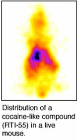
Distribution of a cocaine-like compound in a live mouse
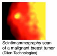
Scintimammography scan of a malignent breast tumor (Dilion Technologies)

