

Preventing Blindness: A Vision of Technology
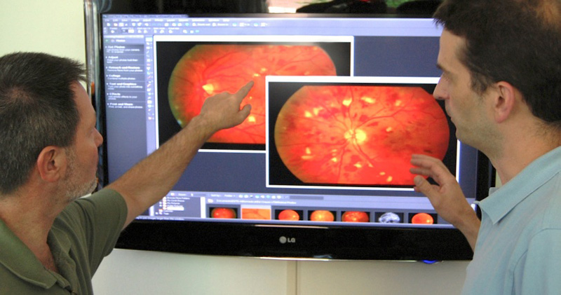
Photo courtesy by Oak Ridge National Laboratory
This project takes advantage of ORNL’s proprietary content-based image retrieval technology, which quickly sorts through large databases and finds visually similar images.
In the blink of an eye, people at risk of becoming blind can be screened for eye diseases such as diabetic retinopathy and age-related macular degeneration. Using a technology originally developed at Oak Ridge National Laboratory (ORNL) to understand semiconductor defects, Automated Medical Diagnostics (AMDx) of Memphis, Tennessee, hopes to establish locations that will allow patients to be screened for eye diseases in their primary care physician’s office and other remote sites.
Why it Matters
Less than half of the 21 million diabetics in the United States receive a recommended annual eye exam, which is critical to minimize serious eye complications and potential blindness. The World Health Organization estimates that by 2025, more than 1 million patients will need to be screened worldwide for diabetes every day. By 2050, the number of diabetics is expected to double.
To reach the goal of screening patients quickly and efficiently, there will have to be a major change in the health care delivery paradigm, but the AMDx Telemedical Retinal Image Analysis and Diagnosis (TRIAD) technology will allow millions of people to undergo screening, especially the indigent and those in medically underserved areas.
Methods
This technology has been used to scan thousands of tiny semiconductors to learn about problems in the manufacturing process for over a decade. The technique adapts this proven technology to describe key retinal regions, which can be used to index images in a content-based image retrieval library. What separates this method from others is the automation of diagnosing retinal disease by capturing ophthalmologists’ knowledge in a patient archive.
Once the physician or technician has a picture of the eyes, the information is sent to a database, where it is compared to thousands of images of known retinal disease states. The system quickly determines whether the patient passes the screening or, if not, provides a follow-up plan that includes visiting an ophthalmologist.
What’s Next
While the plan is to begin with patients in Memphis and the Mississippi Delta, the goal is to have hundreds of cameras throughout the United States and beyond. If diseases can be detected early, treatments can preserve vision and significantly reduce occurrences of blindness. Over the next year, a more fully automated image analysis network to manage images nationwide will be rolled out. Eventually, the goal is to make this a global effort using automated technology and Internet connectivity.
Acknowledgments
This work was led by Ken Tobin of ORNL and Edward Chaum, an ophthalmologist and Plough Foundation professor of retinal diseases at the University of Tennessee Health Science Center Hamilton Eye Institute in Memphis. Other contributors include Tom Karnowski and Luca Giancardo of ORNL, Stacy Li of the UT Health Science Center in Memphis, and Karen Fox of the Delta Health Alliance.
Publications
Giancardo L, F Meriaudeau, TP Karnowski, KW Tobin, P Favaro, A Ruggeri, and E Chaum. 2011. “Textureless Macula Swelling Detection with Multiple Retinal Fundus Images.” IEEE Transactions on Biomedical Engineering 58:3. March.
Giancardo L, F Meriaudeau, TP Karnowski, Y Li, KW Tobin, and E Chaum. 2011. “Quality Enhancement and Nerve Fibre Layer Artefacts in Retina Fundus Images by Off Axis Imaging.” IEEE International Conference on Image Processing, Brussels, Belgium. September.